This award was established by the British Phycological Society in recognition of Hilda Canter-Lund, whose stunning photographs will be known to many members. Her photomicrographs of freshwater algae combined high technical and aesthetic qualities whilst still capturing the quintessence of the organisms she was studying.
Congratulations to all that made the shortlist, the competition was strong this year, and the BPS thanks everyone who submitted images and voted for their favourites.
2026 Winners
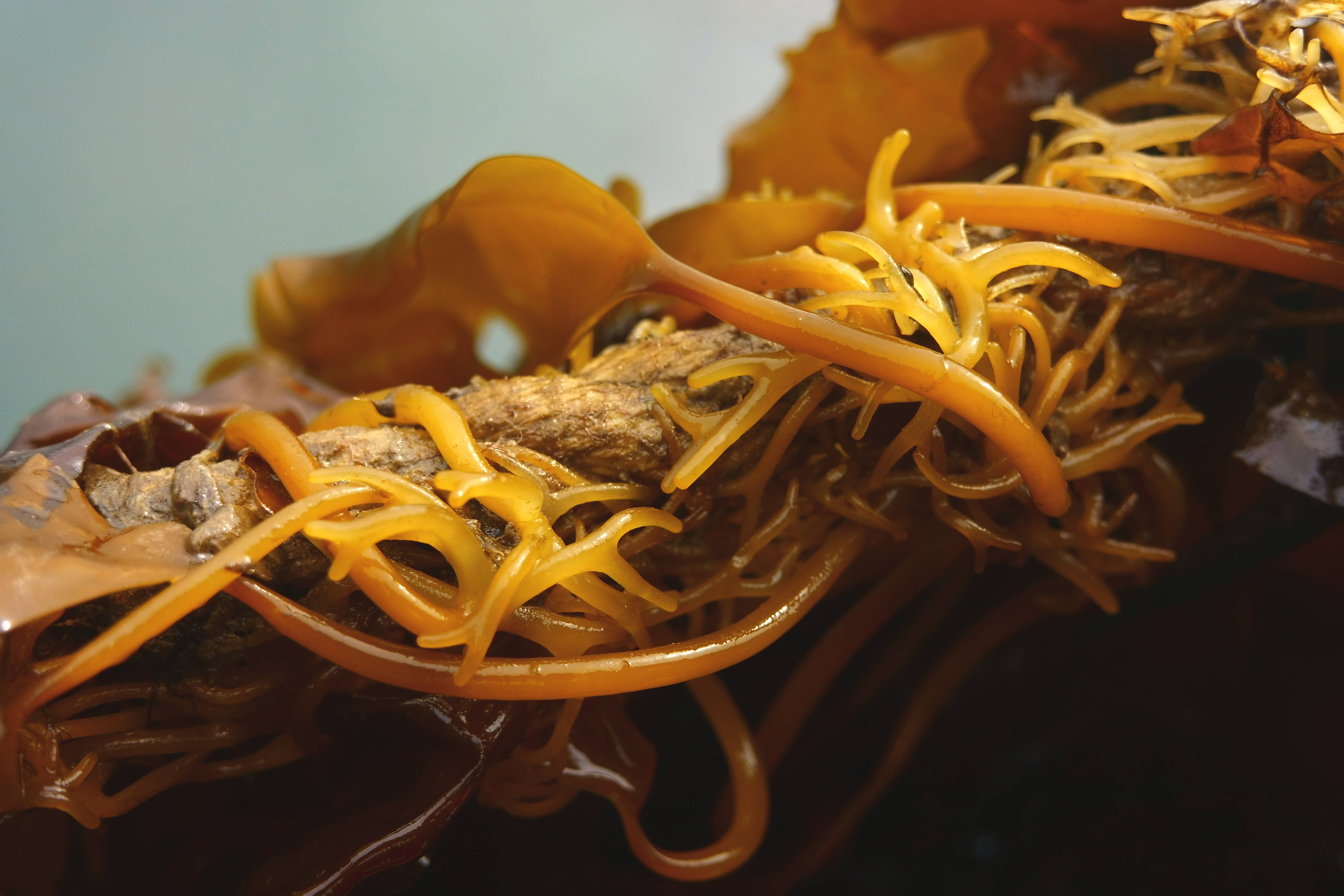
Macro category – Jessica Schiller “Holding Fast”
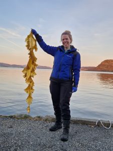
Jessica is a senior scientist at Hortimare in the Netherlands with a lifelong passion for marine biology. Her academic work focused on kelp ecophysiology, specialising in the bioinvasion of Undaria pinnatifida in Europe and its cultivation in Asia during her PhD. As a guest scientist in China, Jessica developed an interest in seaweed cultivation. She moved to the industry, where her work supports seaweed farms, projects and restoration initiatives with seaweed seedlings and research capabilities. For seven years, she has led field trials and an internal research team focused on seaweed attachment, seeding, and farming, combining her academic background with applied research.
Micro category – Joint Dr Jeffery Stone and Hirak Samir Parikh “Geometry of a Glass Cell”
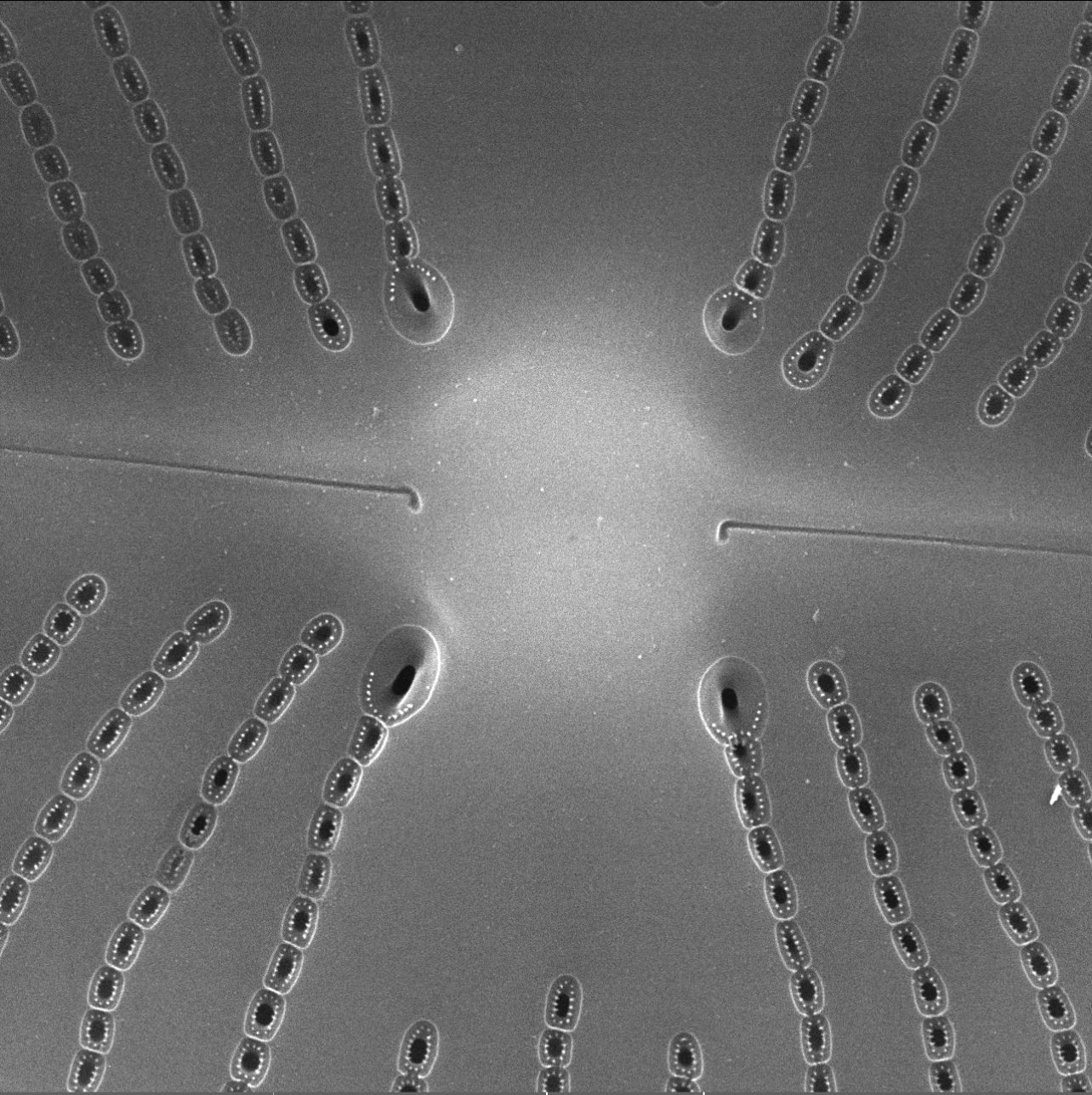
Image originally captured by Dr. Jeffery R. Stone, Indiana State University; edited and refined by Hirak Samir Parikh.
Hirak Samir Parikh is a PhD candidate in the Department of Earth and Environmental Systems at Indiana State University, where his research focuses on diatom taxonomy, systematics, ecology, and their applications in understanding aquatic environmental change. His work combines light and electron microscopy with molecular and ecological approaches to investigate the remarkable diversity, morphology, and evolutionary history of diatoms.
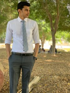
Hirak would also like to acknowledge his academic advisor, Dr. Jeffery R. Stone, who personally captured the original image featured in “Geometry of a Glass Cell.” With Dr. Stone’s permission, Hirak edited and refined the image and submitted it to the competition. Dr. Stone’s mentorship has also been instrumental in developing Hirak’s skills in diatom identification and microscopy and in shaping his appreciation for the intricate valve architecture and microscopic beauty of diatoms.


